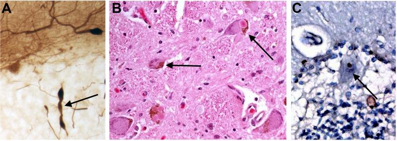
Figure 3.
Three different ET cases showing a heterogeneity of pathological findings in the ETs. A. Representing cerebellar pathology, a Purkinje cell axon is shown with three torpedoes. Calbindin stain. B. Multiple Lewy bodies in the locus ceruleus. Hematoxylin and eosin stain. C. Ubiquitin positive Purkinje cell intranuclear inclusion.

