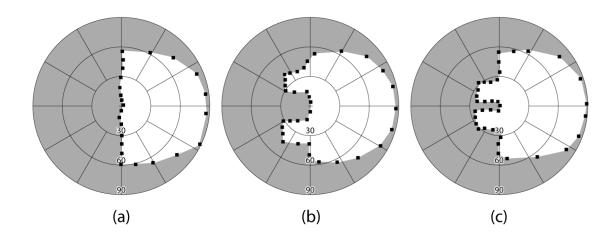
Figure 2. Binocular visual field (Goldmann V4e) of a patient with left HH.
(a) Without peripheral prisms; (b) with 57Δ horizontal peripheral prisms; and (c) with 57Δ oblique peripheral prisms, as fitted for the study with a 12mm inter-prism separation. Both designs provide close to 30° of lateral expansion into the blind hemifield (slightly more for the horizontal than the oblique design). The expansion is in more central areas of the field with the oblique design. Small black squares are the individual points mapped during the perimetry.

