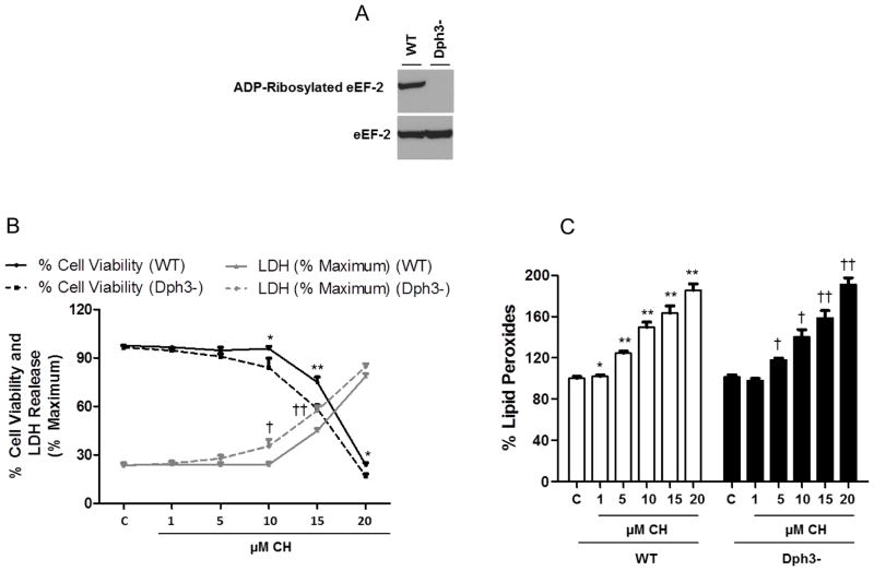
Figure 1.
Diphthamide deficiency increases the vulnerability of cells to oxidative stress. A. Immunoblots for in vitro ADP-ribosylation assay with the cell lysates. The top panel shows ADP-ribosylated eEF-2 and the bottom panel shows the total eEF-2 in wild-type (WT) or diphthamide-deficient cells (Dph3−). B. Cells were incubated for 3 h in absence or presence of the indicated concentrations of CH. Cell viability was measured by the MTS assay and lactate dehydrogenase (LDH) release assay. Values are the mean and SEM of four experiments. *p< 0.05 and **p<0.01 (cell viability), †p<0.05 and ††p<0.01 (cell death); WT versus diphthamide-deficient cells. C. Lipid peroxidation levels were determined using the FOX method. Values are the mean and SEM of 5 independent experiments. *p< 0.05 and **p<0.01 versus the wild-type cell control value. †p<0.05 and ††p<0.01 versus the diphthamide- deficient cell control value.

