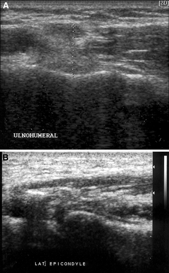
Fig. 2.

a, b Ultrasound (5–12 MHz). Coronal images of lateral epicondyles of two different patients, showing thickened common extensor tendons, loss of the fibrillar echo pattern, and calcific foci (arrows)

a, b Ultrasound (5–12 MHz). Coronal images of lateral epicondyles of two different patients, showing thickened common extensor tendons, loss of the fibrillar echo pattern, and calcific foci (arrows)