Fig. 4.

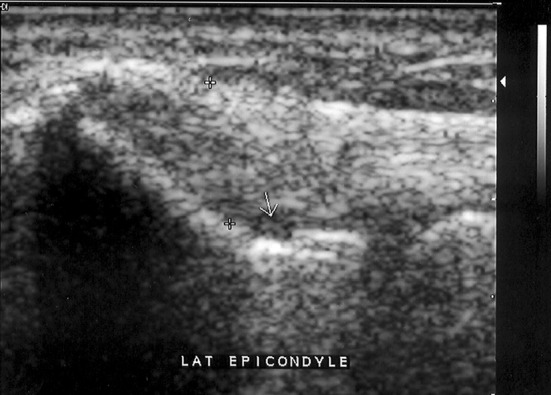
Ultrasound (5–12 MHz). Coronal image of the lateral epicondyle showing focal cortical erosion (arrow). The common extensor tendon also presents fusiform thickening and loss of the fibrillar echo pattern

Ultrasound (5–12 MHz). Coronal image of the lateral epicondyle showing focal cortical erosion (arrow). The common extensor tendon also presents fusiform thickening and loss of the fibrillar echo pattern