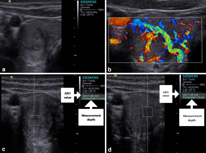
Fig. 3.
Baseline US shows a well marginated, prevalently hyperechoic nodule with hypoechoic capsule (a) which at color-Doppler presented Pattern III and at ARFI 1.89 and 1.56 m/s of shear wave velocity, respectively, for operator 1 (c) and operator 2 (d). Histology after surgery demonstrated that the nodule was benign nodular hyperplasia

