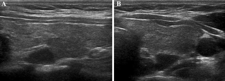
Fig. 3.

Adenoma of the upper left parathyroid gland (middle-superior retrothyroidal, maximum diameter approx. 1.1 cm). Longitudinal (panel A) and axial (panel B) scans. The overlying thyroid parenchyma appears inhomogeneously hypoechoic as a result of chronic autoimmune thyroiditis
