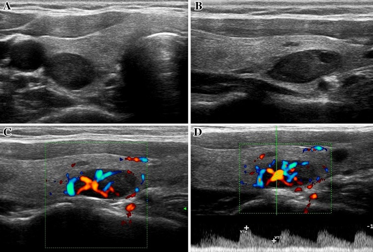
Fig. 7.
Intrathyroidal adenoma of the right inferior parathyroid gland (maximum diameter approx. 1.5 cm circa) that developed within a multinodular goiter. Axial scan (panel A), longitudinal scan (panel B), longitudinal scan with color Doppler imaging (panels C and D). The vascular pattern is predominantly monopolar. Pulsed Doppler sampling of the afferent artery (panel D) revealed typical low-resistance flow (RI 0.52)

