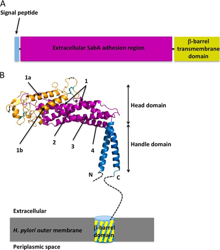
FIGURE 1.
Domain organisation of SabA. A, schematic illustrating the predicted domain structure of SabA. B, crystal structure of SabA N-terminal adhesion region. The handle is in blue, the tetratricopeptide repeat region is colored in magenta and the remainder of the molecule is in orange. Dashed lines indicate disordered regions. The two conserved disulfide bonds are represented as cyan sticks. The short loop interruption in Helix-1 is marked with an asterisk. The C-terminal helix is connected to a trans-membrane β-barrel domain, which anchors the SabA adhesion region to the outer membrane of H. pylori.

