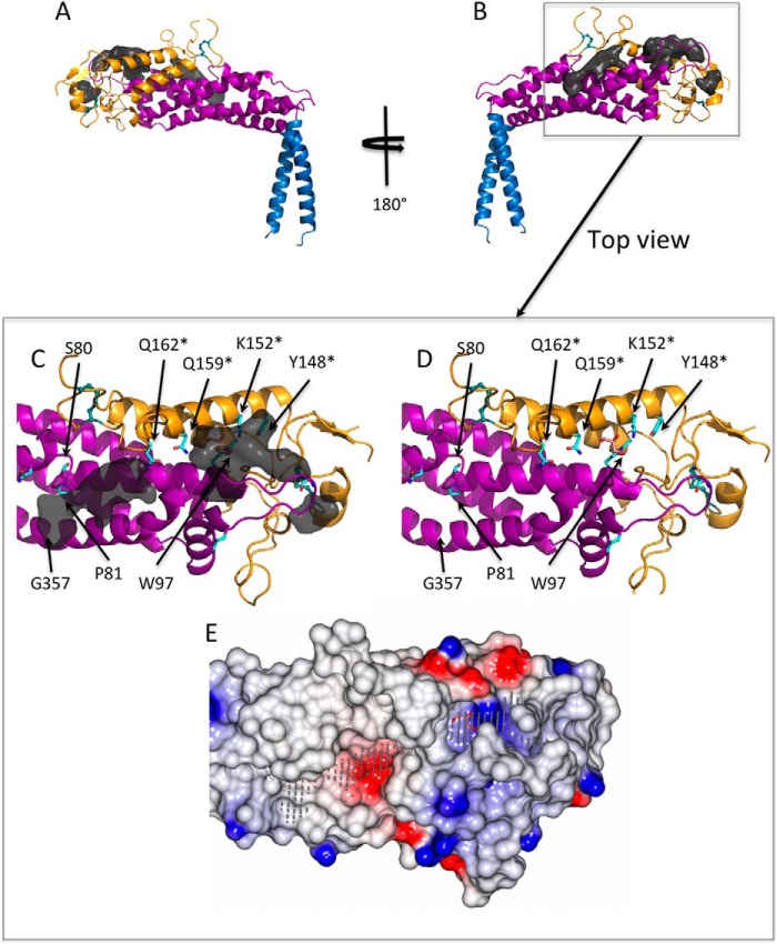
FIGURE 4.
Ligand-binding pockets calculated by an automatic ligand-binding-site prediction program (POCASA). A and B, the binding pockets (dark gray surface) are predicted mostly toward to the tip of the SabA head domain. C and D, view from the top toward the predicted ligand binding pockets (removed in D for clarity). A number of highly conserved residues (cyan sticks), in particular Ser-80, Pro-81, Trp-97, Tyr-148, Lys-152, Gln-159, and Gln-162, line the surface of these pockets. Tyr-148, Lys-152, Gln-159, and Gln-162 were mutated to alanine in this study are marked with an asterisk. A glycerol molecule (pink sticks) is observed in the crystal structure and forms hydrogen bond interactions with Lys-152 and Gln-159. E, electrostatic potential surface representation of the SabA head domain showing the predicted pockets (gray dots) generated using CCP4 mg (30). A glycerol molecule (pink sticks) binds in a positively charged cavity.

