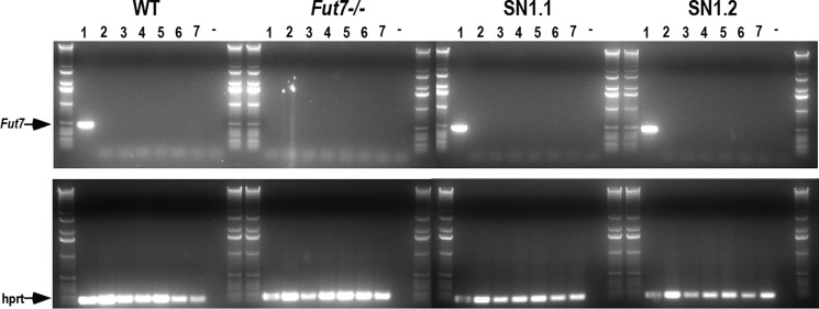
FIGURE 6.

Absence of Fut7 expression in inappropriate cell types. Mice were anesthetized, perfused with PBS, sacrificed, and total RNA was isolated and reverse-transcribed, and PCR was performed on organs from WT, Fut7 KO, SN1.1, and SN1.2 mice. Lanes represent RT-PCR from bone marrow (1), brain (2), lung (3), liver (4), kidney (5), skeletal muscle (6), and small intestine (7). The dash (−) represents negative control of no cDNA input to the PCR reaction. Primers for hprt were used as a positive control on all samples (lower panels). The entire PCR reaction was run on 1% agarose gels, with each set of eight samples flanked by DNA size ladders.
