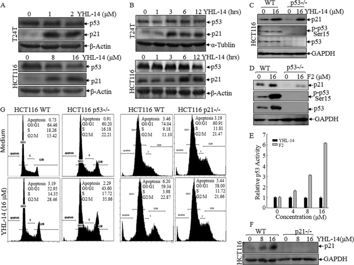
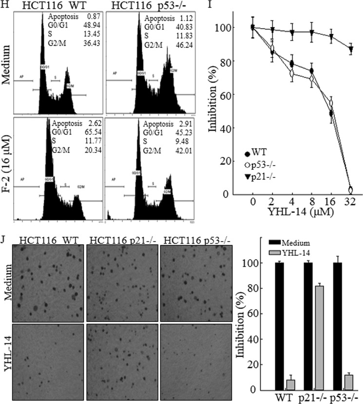
FIGURE 3.
p21 protein expression up-regulated by YHL-14 plays a key role in the induction of G2/M phase growth arrest in cancer cells. A and B, after synchronization, cells were treated with YHL-14 at the indicated concentrations for 12 h (A) or YHL-14 at 2 μm (T24T cells) or 16 μm (HCT116 cells) for the indicated time points (B). C and D, HCT116 WT, HCT116 p53−/−, and HCT116 p21−/− cells were treated with the indicated doses of YHL-14 (C) or F2 (D) for 12 h. The cell extracts were subjected to Western blotting with anti-p21, anti-p53, anti-phospho-p53 (Ser-15), or anti-GAPDH antibodies. E, HCT116 stable transfectant (8 × 10³) that was stably transfected with the PG13-luciferase reporter was seeded into each well of a 96-well plate. After synchronization, cells were treated with the indicated concentrations of YHL-14 or F2 for 12 h, and the cells were then extracted for determination of luciferase activity. The results were presented as p53-dependent luciferase transactivity relative to vehicle control (relative p53 activity). Error bars show the mean ± S.D. from three independent experiments. F, HCT116 WT and HCT116 p21−/− cells were treated with YHL-14 at concentrations as indicated for 12 h, and the cell extracts were subjected to Western blotting with anti-p21 and anti-GAPDH antibodies. G and H, HCT116 WT, HCT116 p53−/−, and HCT116 p21−/− cells were treated with the indicated doses of YHL-14 (G) or F2 (H) for 12 h. The cells were then fixed and subjected to flow cytometry analysis. I, results of a coupled ATPase activity assay in the presence of varying concentrations of YHL-14 at 24 h. Incubation with YHL-14 caused dose-dependent growth effects of HCT116 wild-type, HCT116 p21−/−, and HCT116 p53−/− cells in vitro as observed in ATPase assays. Proliferation rates were determined in the indicated cells using a CellTiter-Glo luminescent cell viability assay kit. Results are presented as the mean ± S.D. of triplicate assays. J, HCT116 WT, p21−/−, and p53−/− cells were seeded in soft agar as described under “Experimental Procedures.” Representative images of colonies of these cells in a soft agar assay without or with YHL-14 (16 μm) were visualized under the microscope, and only colonies with more than 32 cells were counted. Colonies are expressed as mean ± S.D. from five assays of three independent experiments.


