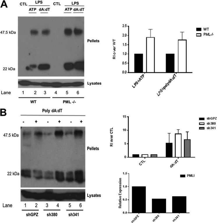
FIGURE 2.
Loss of PML enhances NLRP3- and AIM2-stimulated ASC oligomerization. A, left panel, primary BMDM from 129Sv wild type (WT) and PML-deficient mice were left unstimulated or stimulated with 100 ng/ml LPS for 3 h with or without the addition of 5 mm ATP for 1 h or transfected with 1 μg/ml poly(dA-dT) for 5 h as indicated. Cell pellets were washed and resuspended in PBS and then cross-linked by incubation with 20 mm DSS for 30 min. Cross-linked pellets and cell lysates were analyzed by Western blotting with anti-ASC antibody (AL177). Right panel, densitometry of oligomerization experiments is presented. Each dimer band was normalized to whole cell lysate ASC content, and the relative intensity (RI) of the dimer band (47.5 kDa) over the whole cell lysate (set at 1) was calculated. The data shown are representative of three independent experiments. CTL, control. B, left panel, all three THP-1 cell lines were differentiated with 12-myristate 13-acetate for 16 h. Following this, cells were stimulated with 2 μg/ml poly(dA-dT) for 5 h. Cell lysates were centrifuged at 330 × g for 10 min at 4 °C. Cell pellets were again analyzed for ASC by Western blotting as in A. Right panel, top row, densitometry of oligomerization in THP-1 cells lines with stable knockdown of PML. Each band was normalized to its corresponding whole cell lysate ASC content, and the relative intensity (RI) of the dimer band (47.5 kDa) over the whole cell lysate (set at 1) was calculated. The data shown are representative of two independent experiments. Right panel, bottom row, mRNA knockdown of PMLI in THP-1 cell lines generated using two shRNA lentiviral vectors (sh380 and sh341). Nonsilencing lentiviral vector pGIPZ was used for generation of nontargeting control cell line, shGPZ.

