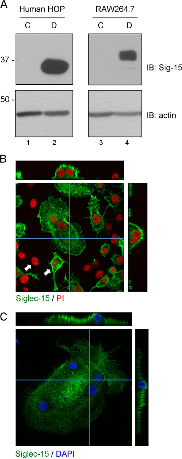
FIGURE 1.

Expression and localization of Siglec-15 protein in osteoclasts. A, Western blots of total protein extracts from human HOP- and mouse RAW264.7-derived osteoclasts (D) and non-differentiated precursor cells (C). B and C, confocal microscopy analysis of Siglec-15 localization in RAW264.7- and HOP-derived osteoclasts. B, RAW264.7 cells were grown for 3 days on glass coverslips in the presence of RANKL. Fixed and permeablized cells were then stained with anti-Siglec-15 and propidium iodide (PI). Z-stacks were prepared by acquiring confocal images at 1-μm intervals throughout the depth of the cells in the field. The central panel shows a single confocal image of a multinucleated osteoclast surrounded by non-fused precursor cells. The two arrows indicate examples of mononuclear cells that are positive or negative for Siglec-15 expression. The narrow panels on top and right are cross-sections through the osteoclast generated by re-slicing a three-dimensional reconstruction prepared from the image stack (the slices were performed along the paths indicated by the blue horizontal and vertical lines). C, human HOP-derived osteoclasts were grown on chamber slides, fixed, and stained with anti-Siglec-15 and DAPI. Analysis by confocal microscopy was performed as described in B.
