FIGURE 3.
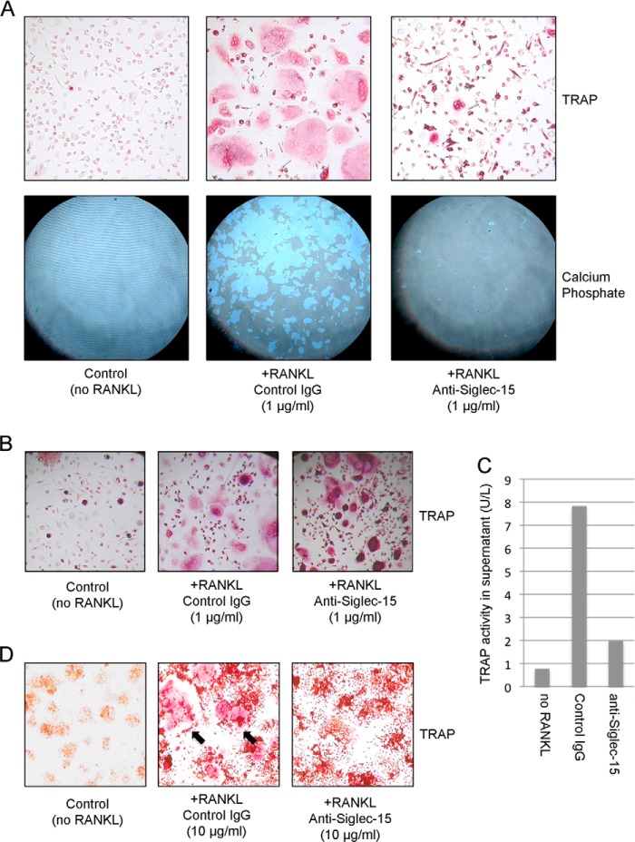
Siglec-15 antibodies inhibit osteoclast differentiation and activity in vitro. A, HOPs were grown with macrophage colony-stimulating factor alone (left panels) or with macrophage colony-stimulating factor and RANKL (to induce osteoclast differentiation) in the presence of control human IgG (middle panels) or anti-Siglec-15 (right panels) for 7 days. Cells were grown in parallel either on plastic (top panels) or on calcium phosphate-coated (bottom panels) plates. Multinucleated osteoclasts were identified on plastic by TRAP staining (red). Regions of calcium phosphate resorption were visualized by phase-contrast mode light microscopy after cell removal. B, upon prolonged treatment with RANKL (10 days rather than 7 days), HOPs grown in the presence of anti-Siglec-15 E09 fuse to form intensely TRAP-stained, multinucleated cells (right panel). The morphology of these cells is distinct from normal osteoclasts formed in the presence of a control antibody (middle panel). C, HOPs differentiated with RANKL in the presence of anti-Siglec-15 E09 display impaired TRAP secretion. TRAP levels in conditioned media of HOPs differentiated for 10 days were determined by ELISA. D, Siglec-15 antibody B02 inhibits differentiation of mouse RAW264.7 cells into osteoclasts. RAW264.7 cells were treated with RANKL to induce differentiation in the presence of control human IgG (middle panel) or anti-Siglec-15 B02 (right panel). Control cells were grown without RANKL or antibodies (left panel). After 3 days in culture, cells were stained for TRAP activity (red). The black arrows in the middle panel indicate examples of multinuclear, TRAP-positive osteoclasts.

