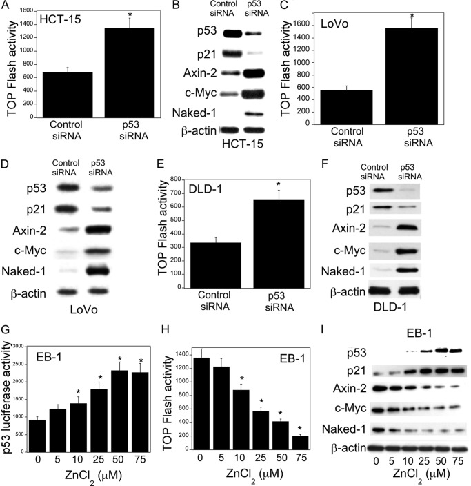
FIGURE 1.
p53 regulates Wnt signaling. A, C, and E, cells were transfected with 0.45 μg each of TOP Flash and FOP Flash constructs and 0.2 μg of pSVβgal. Cells also received 0.9 μg of siRNA to GFP (control siRNA) or p53. 48 h after transfection, cells were harvested, and luciferase activity was measured. TOP Flash activity was determined by the ratio of pTOP-flash to pFOP-flash luciferase activity, each normalized to β-galactosidase enzymatic activity levels. B, D, and F, cells were transfected with 2 μg of siRNA (control) to GFP or p53 for 48 h. Following transfection, cells were harvested, and cell lysates were subjected to Western blotting. The blots were probed with antibodies to the indicated proteins. G and H, EB-1 cells were transfected with 1.8 μg of p53 luciferase construct (G) or 0.9 μg each of TOP Flash and FOP Flash constructs and 0.2 μg of pSVβgal for 24 h. 24 h later, cells were treated with indicated concentrations of ZnCl2 for 12 h, and then cells were harvested, and luciferase activity was measured. Luciferase activity was normalized to β-galactosidase activity. I, cells were treated with the indicated concentrations of ZnCl2 for 12 h. Cell lysates were subjected to Western blotting, and the blots were probed as indicated. A, C, E, G, and H, mean ± S.D. (error bars) are shown, n = 6. *, p < 0.01 compared with control siRNA-treated cells (A, C, and E) or vehicle-treated cells (G and H).

