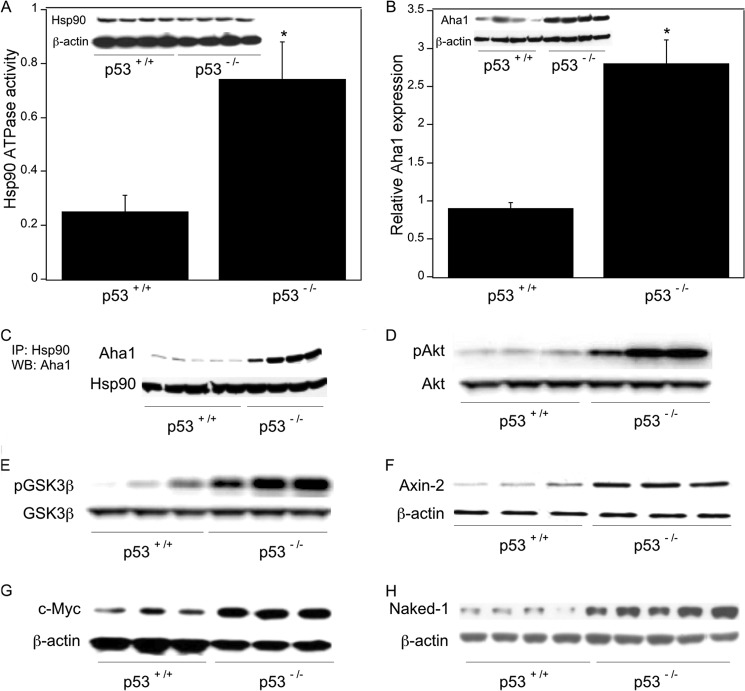
FIGURE 9.
Wnt signaling is activated in p53-null mice. Colon tissue from p53 wild type and p53-null mice was used. Poly(A) RNA was isolated from total RNA extracted from colon tissues. Additionally, tissues were homogenized, and protein lysates were prepared. A, tissue lysates were used to measure Hsp90 ATPase activity using a protocol described under “Experimental Procedures.” Mean ± S.D. (error bars) are shown, n = 6. *, p < 0.01 compared with p53+/+ mice. Lysates were also subjected to immunoprecipitation with Hsp90 or β-actin antibodies; Western blotting was carried out, and the blot was probed for Hsp90 and β-actin as indicated. B, relative expression of Aha1 was quantified by real-time PCR. Values were normalized to levels of β-actin. Mean ± S.D. are shown, n = 6. *, p < 0.01 compared with p53+/+ mice. Tissue lysates were also immunoprecipitated with Aha1 or β-actin antibody; Western blotting was carried out, and the blots were probed for Aha1 and β-actin as indicated. C, tissue lysates were immunoprecipitated (IP) with Hsp90 antibody, and the immunoprecipitates were subjected to Western blotting (WB) for Aha1 and Hsp90 as indicated. D and E, tissue lysates were immunoprecipitated with antibodies to Akt (D) or GSK3β (E), and the blots were probed for pAkt and Akt (D) or pGSK3β and GSK3β (E) as indicated. F–H, tissue lysates were immunoprecipitated with Axin-2, c-Myc, Naked-1, or β-actin antibodies, and the blots were probed with the same antibodies.

