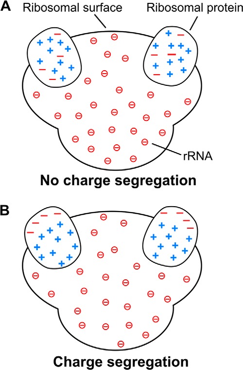
FIGURE 4.

Schematic models for the charge distribution of ribosomal proteins illustrating the charge segregation concept. A, ribosomal proteins with no intramolecular charge segregation. B, ribosomal proteins with intramolecular charge segregation supported by this study. The negative charges enclosed in circles are from the phosphate groups of rRNA. For simplicity, only two ribosomal proteins are shown embedded in each ribosome.
