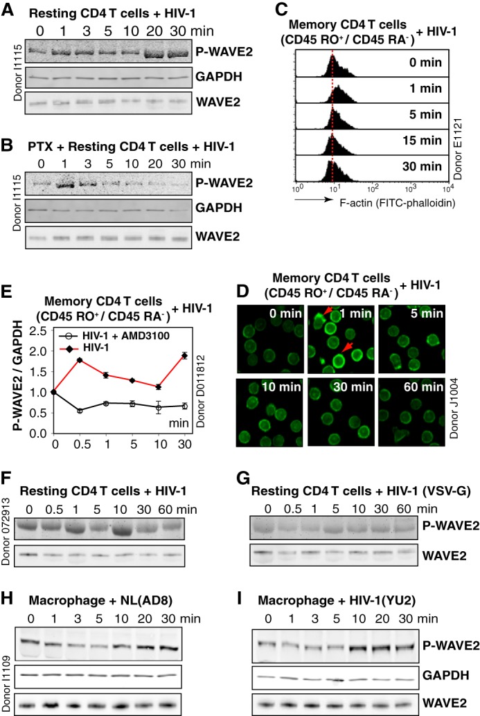
FIGURE 1.

HIV-1 triggers WAVE2 serine 351 phosphorylation. A, resting CD4 T cells were treated with HIV-1NL4–3 over the indicated time course, and WAVE2 serine 351 phosphorylation was detected by an IR fluorophore-conjugated, anti-phospho-S351 WAVE2 antibody. The same blot was also stained with an anti-human GAPDH antibody for loading control. WAVE2 was also stained with an anti-WAVE2 antibody on a blot duplicate. B, resting CD4 T cells were treated with 50 ng/ml PTX, similarly treated with HIV-1, and analyzed for WAVE2 S351 phosphorylation. C and D, resting memory CD4 T cells (CD45RO+/CD45RA−) were treated with HIV-1 for a time course, and F-actin was stained with FITC-phalloidin and analyzed with flow cytometry (C) or confocal microscopy (D). Red arrows indicate localized actin polymerization. E, resting memory CD4 T cells were pre-treated or not with AMD3100 (100 nm), and then infected with HIV-1 for a time course. WAVE2 serine 351 phosphorylation was similarly analyzed and quantified. F and G, resting CD4 T cells were treated with HIV-1NL4–3 or with HIV-1(VSV-G), and WAVE2 serine 351 phosphorylation was measured. H and I, blood monocyte-derived macrophages were treated with HIV-1NL4–3 (AD8) or HIV-1YU2 for a time course, and WAVE2 serine 351 phosphorylation was similarly analyzed.
