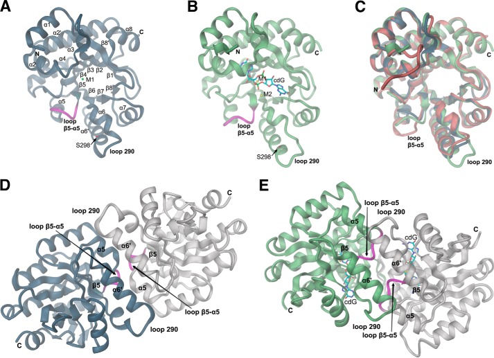
FIGURE 1.
Crystal structures of the YahA-EAL domain. Schematic representation with secondary structure elements and chain termini labeled. The β5-α5 loop is highlighted in magenta. A, structure of the monomer of YahA-EAL in complex with Mg2+ (M1). Mutation site Ser-298 is shown in full and labeled. B, structure of the monomer of YahA-EAL in complex with substrate (cdG) and Ca2+ (M1 and M2). C, superimposition of YahA-EAL-apo (brown) with YahA-EAL·Mg2+ (steel blue) and YahA-EAL·cdG·Ca2+ (light green). D, subunit arrangement in YahA-EAL·Mg2+ (canonical EAL dimer). The asymmetric unit contains two dimers, both are virtually identical. E, subunit arrangement in YahA-EAL·cdG·Ca2+ (closed EAL dimer). The asymmetric unit contains one dimer with virtually identical subunit structure. In panels D and E the same color code is used as in panels A and B, but with symmetry mates shown in gray.

