
FIGURE 5.
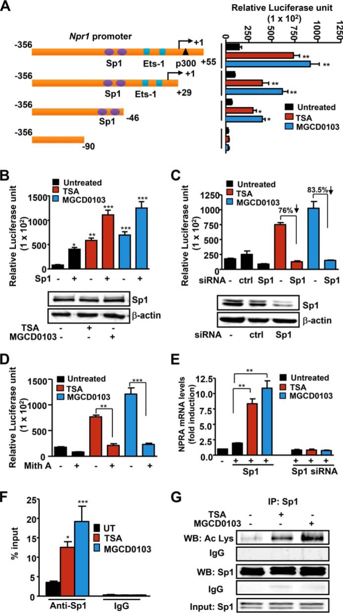
Role of Sp1 in mediating TSA and MGCD0103 effects on Npr1 gene transcription. A, schematic map of the Npr1 promoter deletion construct and their luciferase activity in transfected MMCs treated with HDACi. B, luciferase activity of the Npr1 promoter construct −356/+55 cotransfected with Sp1 plasmid and treated with HDACi. Blots show Western blot analysis of Sp1 protein expression in HDACi-treated cells with β-actin as loading control. C, luciferase activity of the Npr1 promoter cotransfected with Sp1 siRNA and treated with HDACi. Lower panel, Western blot analysis of Sp1 protein expression in Sp1 siRNA-transfected cells and β-actin as loading control. D, luciferase activity of the Npr1 promoter in cells pretreated with mithramycin A and induced with HDACi. E, effect of HDACi on Npr1 mRNA levels in cells transfected with Sp1 expression plasmid or Sp1 siRNA as determined by real-time RT-PCR with β-actin as the internal control. F, quantitative ChIP assay demonstrating recruitment of Sp1 protein on the Npr1 promoter (−120 to +73) in HDACi-stimulated cells as determined by real-time PCR. G, Western blot analysis of acetylated and total Sp1 in the immunoprecipitate from HDACi-treated cells. Input shows Sp1 in lysates as detected by Western blot. Bars represent mean ± S.E. of three independent experiments. HDACi (TSA, 50 nm; MGCD0103, 1 μm); Ctrl, control; UT, untreated; mith A, mithramycin A; WB, Western blot; IP, immunoprecipitation; *, p < 0.05; **, p < 0.01; ***, p < 0.001.