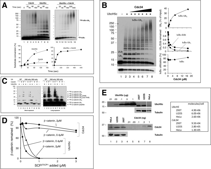
FIGURE 2.
In vitro ubiquitination of IκBα and β-Catenin by SCFβTrCP2, UbcH5c, and Cdc34. A, kinetic analysis of polyubiquitination of IκBα-(1–54) by SCFβTrCP2 in the presence of UbcH5c, Cdc34, or both UbcH5c and Cdc34. Ubiquitination was carried out as described under “Experimental Procedures” with substrate (0.1 μm), E3 (0.1 μm), UbcH5c (17 μm), and Cdc34 (17 μm). B, effect of Cdc34 concentration on polyubiquitination of IκBα-(1–54) by SCFβTrCP2 in the presence of UbcH5c. Ubiquitination was carried out as in panel A at 37 °C for 3 min. Reaction products are marked on the gel. Although Ub1 denotes mono-ubiquitinated IκBα-(1–54) at either Lys-21 or Lys-22, 2Ub represents the di-Ub products with Ub conjugated at both Lys-21 and Lys-22. * refers to IκBα-(1–54) attached with a di-Ub chain formed at either Lys-21 or Lys-22. C, combined actions of UbcH5c and Cdc34 drive polyubiquitination of β-catenin by SCFβTrCP2. Ubiquitination was carried out as in panel A with the wild type Ub or variants as indicated at 37 °C for 15 min. Both short and long exposures are shown. Substrate and reaction products are marked on the gel, with numbers denoting the number of Ub moieties. D, analysis of polyubiquitination of β-Catenin by SCFβTrCP1 with UbcH5c or Cdc34 in a range of concentrations of substrate or E3. Varying concentrations of β-catenin or SCFβTrCP1, as indicated, were used for the ubiquitination assay. The results were quantified and are shown graphically. E, immunoblot analysis of cellular concentrations of UbcH5 and Cdc34. Protein extracts from the indicated cell lines were subject to immunoblot analysis along with purified UbcH5c and Cdc34 in amounts as indicated. For the immunoblots of UbcH5 or Cdc34, protein lysates derived from 300,000 or 150,000 cells, respectively, were used. The box indicates the estimated number of molecules for UbcH5 or Cdc34 in the indicated cell line.

