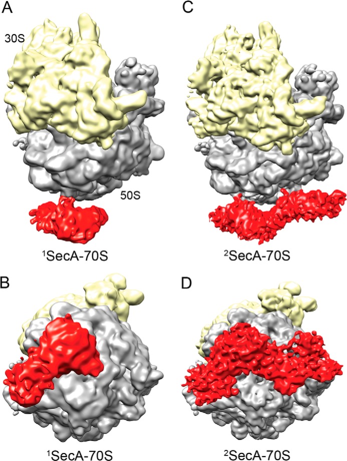
FIGURE 3.

Cryo-EM reconstructions of SecA bound to the 70S ribosome. A, cryo-EM structure of monomeric SecA bound to the 70S ribosome (1SecA-70S). The 30S small ribosomal subunit and the 50S large subunit are shown in yellow and gray, respectively. Additional density at the tunnel exit site, shown in red, represents monomeric SecA. B, the same as A but rotated upwards by 90° around the horizontal axis. C, cryo-EM structure of two copies of SecA bound to the 70S ribosome (2SecA-70S). The density at the tunnel exit site represents two copies of SecA. The ribosomal subunits and SecA are shown in the same colors as in A. D, same as C but rotated upwards by 90° around the horizontal axis.
