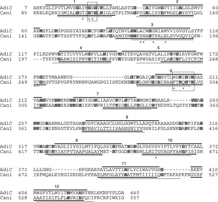
FIGURE 1.
Alignment of Can1 and AdiC sequences. The TM helices as determined by STRIDE (67) on the crystal structure of the OF occluded AdiC state (PDB code 3L1L (8)) are shown as black lines above the AdiC sequence. The TM helices predicted on the Can1 sequence by either HMMTOP (black) (28) or TMPRED (gray) (29) are shown as solid lines below the Can1 sequence. Residue motifs (see “Results”) are framed, and residues interacting with the substrate (see Table 4) are marked by an asterisk. Conserved residues in AdiC and Can1 are boxed in gray.

