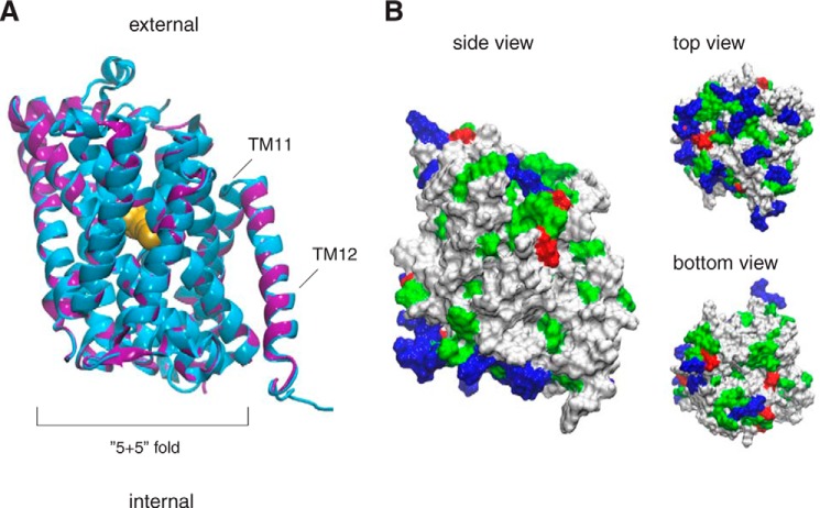
FIGURE 2.
Three-dimensional model of Can1 permease. A, best energy-scored three-dimensional model of Can1 in the OF occluded state superimposed onto the AdiC crystal structure (PDB code 3L1L) (8). The last two TMs (TM11 and TM12) are not part of the “5 + 5” fold. The proteins are depicted in a ribbon diagram, with AdiC in blue and Can1 in purple. The arginine ligand of the AdiC crystal structure is shown as yellow spheres. The two long extracellular loops located in Can1 between TM5 and TM6 and between TM7 and TM8 are not shown. B, three views of the molecular surface of the Can1 model colored according to the residue type (hydrophobic in white, polar in green, positively charged in blue, and negatively charged in red). The figure was prepared with vmd (68).

