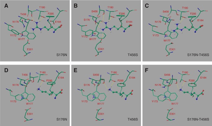
FIGURE 7.
The binding mode of arginine and of lysine predicted in the Can1 modeled three-dimensional structure. Arginine bound in Can1(S176N) variant featuring one Asn-176 rotamer (A), Can1(T456S) variant featuring one Ser-456 rotamer (B), and Can1(S176N-T456S) variant featuring one Asn-176 and one Ser-456 rotamer (C). Lysine bound in Can1(S176N) variant featuring one Asn-176 rotamer (D), Can1(T456S) variant featuring one Ser-456 rotamer (E), and Can1(S176N/T456S) variant featuring one Asn-176 and one Ser-456 rotamer (F). Arginine and lysine are depicted as ball-and-stick representations, and surrounding residues are shown as sticks. Residues are colored according the following scheme. Green, carbon; red, oxygen; blue, nitrogen; white, hydrogen. Hydrogen bonds are shown as yellow broken lines.

