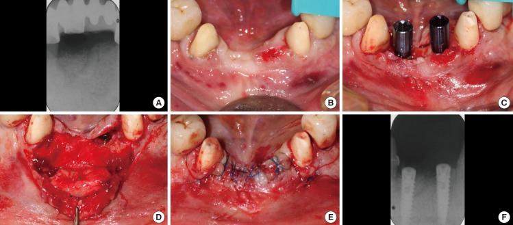
Figure 7.
(A) Periapical radiograph taken 5 months postoperation. (B) Before the implant placement surgery and 5 months post implantotomy. (C) Osteotomies were prepared and the implants were placed centrally on the crestal bone in positions 41 and 31 (FDI-Notation), both with an initial seating torque of 50 Ncm; the final seating was carried out with a hand wrench at >50 Ncm. (D) Position 41 has been further 'grafted' with Bio-Oss granules and covered with its membrane to enhance the hard tissue volume at the site. Connective tissue was harvested from the palate and sutured to the underside of the mucoperiosteal flap using 6.0 Vicryl Rapide to prevent its exfoliation. (E) Wound closure via the coronal advancement of the labial flap using 5.0 Prolene sutures. (F) Periapical radiograph taken immediately postoperatively.

