
Figure 3. Colocalization of antiquitin with the astrocyte marker GFAP.
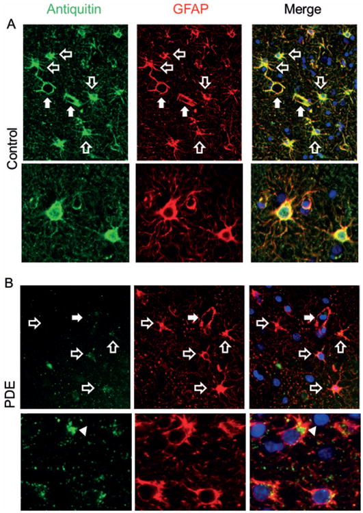
Antiquitin (green) and GFAP (red) immunofluorescence in Control (A) and PDE (B) cortex. In control brain, the distribution of antiquitin and GFAP overlaps. In PDE cortex, antiquitin immunofluorescence is faint and in some astrocytes is accumulated near the nucleus (arrowhead). Open arrows indicate astrocytes; closed arrows indicate small cortical blood vessels, which are surrounded by strong antiquitin immunoreactivity in control, but not PDE, cortex. Blue = nuclear counterstain DAPI.