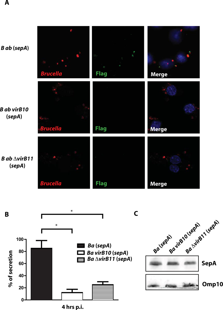
Figure 3. Secretion of SepA is a virB dependent process.
A. Representative Immunofluorescence of J774 A.1 cells infected with B. abortus wild type, virB10 and ΔvirB11 mutant strains coding for SepA-3xFLAG in a replicative vector at 4 hrs post-infection. Red, Brucella; green, FLAG. B. Quantification of the percent of SepA secreting bacteria of the representative images shown in A. *P<0.05. C. Western-blot with an anti-FLAG of the strains showing equivalent levels of expression of SepA.

