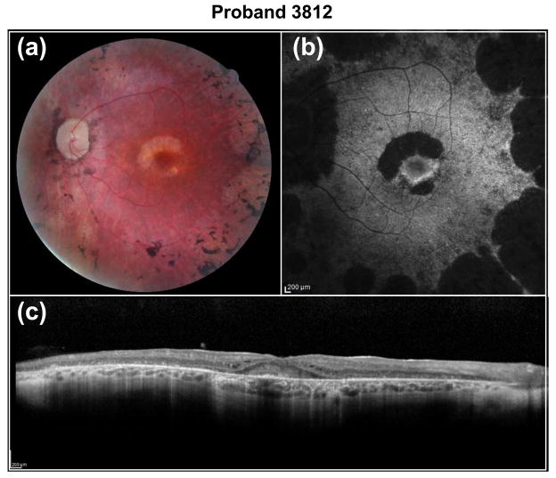
Fig. 4.
Fundus images of proband 3812. Shown are (a) fundus photograph, (b) fundus autofluorescence (FAF) and (c) optical coherence tomography (OCT) images. The images of OS reveal an extensive maculopathy in a horsehoe pattern, with absent FAF pericentrally and marked retinal remodelling with extensive debris and CME in the fovea

