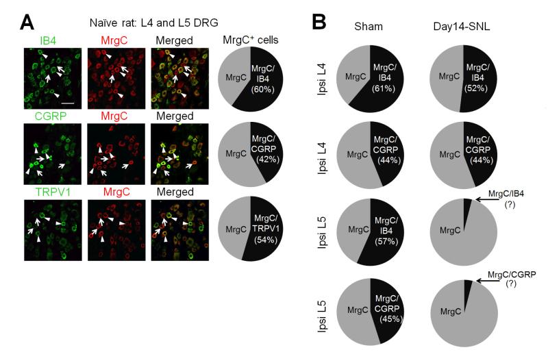
Fig. 4.
(A) Left: Double-immunofluorescence staining shows the distribution of MrgC immunoreactivity (+) in different subsets (IB4, CGRP) of lumbar DRG neurons in naïve rats. MrgC+ neurons that coexpressed other molecules are indicated by arrowheads, and MrgC+ neurons negative for other molecules are indicated by arrows. Right: The proportions of double-labeled cells in MrgC+ neurons in lumbar DRGs of naïve rats (n=3). Scale bar: 50 μm. (B) The proportions of double-labeled cells in MrgC+ neurons in ipsilateral (Ipsi) L4 and L5 DRGs of rats on day 14 post-SNL (n=3) and after sham-surgery (n=2).

