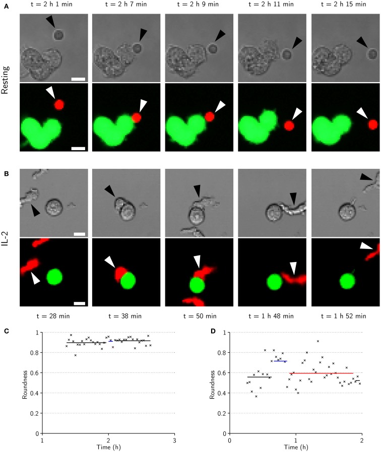
Figure 6.
IL-2-activated NK cells are larger, have elongated shapes during migration, and spread across the target cell during conjugation. (A) Time-lapse sequence showing transmitted-light (top) and fluorescence (bottom) images of a resting NK cell (arrowheads) approaching (t = 2 h 1 min) a cluster of three target cells (green in fluorescence images). The NK cell is briefly conjugated to the target cell (t = 2 h 7 min), before it appears to end its commitment to the target and enter an attachment phase (t = 2 h 9 min) that ends as the NK cell detaches (t = 2 h 11 min) and resumes free migration (t = 2 h 15 min). Scale bars indicate 10 μm. (B) Same as in (A) but for an activated NK cell. The NK cell approaches a target cell (t = 28 min) and forms a conjugate (t = 38 min) that ends as the NK cell assumes a migratory morphology (t = 50 min). The NK cell remains attached to the target cell for almost an hour before it detaches (t = 1 h 48 min) and migrates away from the target cell (t = 1 h 52 min). (C) NK cell roundness vs. time for the sequence shown in (A). (D) NK cell roundness vs. time for sequence shown in (B). Shown are also mean roundness in conjugation (blue lines), attachment (red lines), and free migration (black lines).

