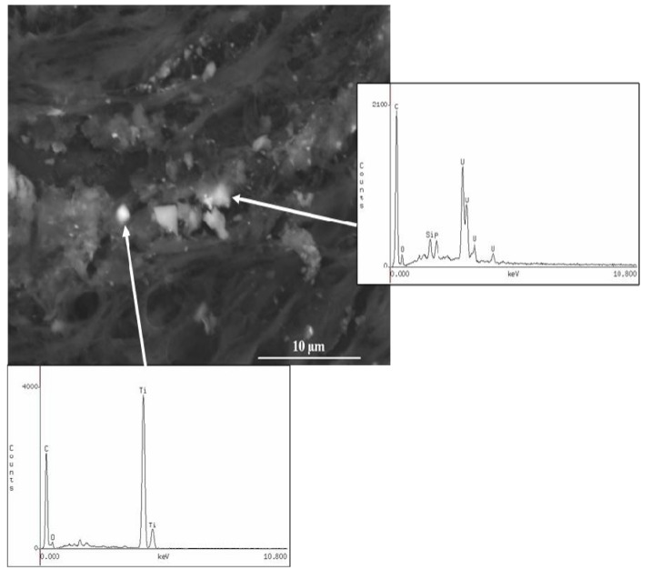
Figure 5.
Case 2: Scanning electron microscopy (SEM) image of the DU fragment and energy-dispersive X-ray analysis (EDXA) of a region of tissue with granular black fragments demonstrating the presence of uranium and titanium. Depleted Uranium particles were measured in in the range of 0.8–1.69 µm. SEM original magnification 3,000×.

