Figure 2.

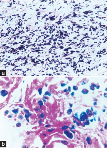
(a) Black-pigmented expectoration. Cell block section (H and E, ×200). (b) Black-pigmented expectoration. Cell block section (Pearl's stain, ×400)

(a) Black-pigmented expectoration. Cell block section (H and E, ×200). (b) Black-pigmented expectoration. Cell block section (Pearl's stain, ×400)