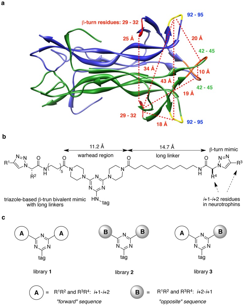
Figure 2. Relative structure of mimetics within neurotrophin proteins.
(a) Distances between hot-spots (highlighted with red, orange, and yellow) in NT-3. (b) General structures of triazole-based β-turn mimics with long linkers, and lengths of warhead region and linker. (c) Strategy to build bivalent mimics with different orientation sequences in triazole core.

