Figure 1.
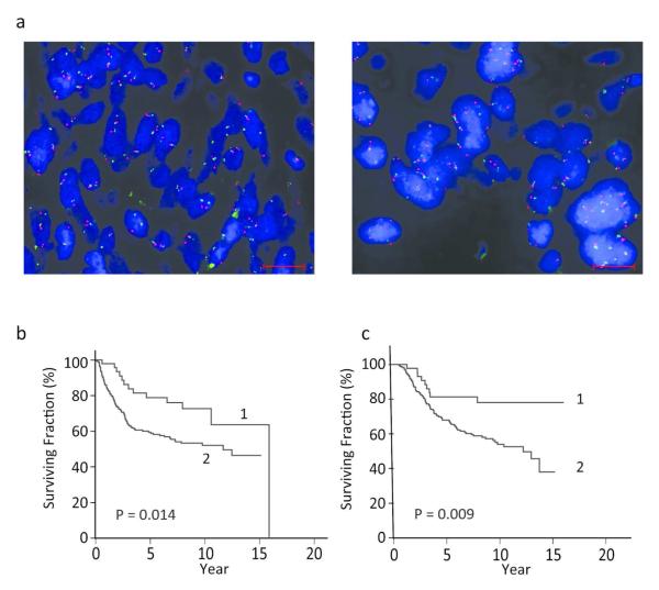
PHIP copy number and its correlation with DMFS and DSS in melanoma patients. (a) Dual color FISH for PHIP locus, red, and centromere of chromosome 6, green, representative of low PHIP copy number, left panel, and high copy number, right panel. Scale bar = 20 Rm. (b) Kaplan-Meier analysis of DMFS in patients with low PHIP copy number (curve 1) versus patients with high PHIP copy number (curve 2). (c) Kaplan-Meier analysis of DSS in patients with low PHIP copy number (curve 1) versus patients with high PHIP copy number (curve 2).

