Figure 2.
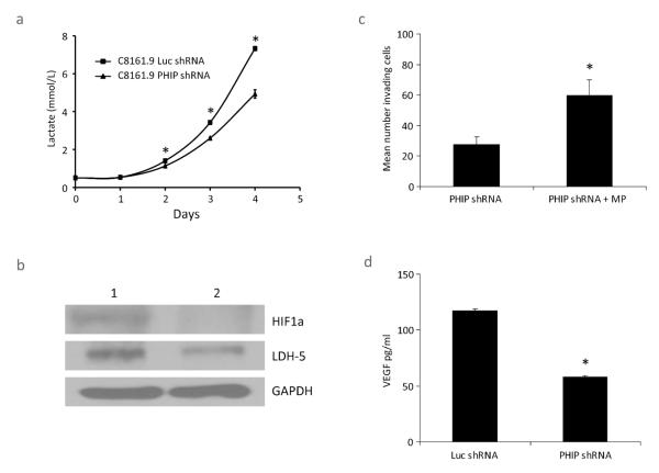
Effects of shRNA-mediated suppression of PHIP in human melanoma cells. (a) Lactate production in C8161.9 cells expressing anti-PHIP shRNA versus control cells expressing anti-luc shRNA. Mean ± SE; N = 3. * denotes P < 0.003. (b) Western analysis of LDH5 and HIF1A in C8161.9 cells expressing anti-luc shRNA (control cells, lane 1) or anti-PHIP shRNA (lane 2). (c) Invasive capacity of C8161.9 stable transformants expressing anti-PHIP shRNA with or without the addition of methylpyruvate (MP). Mean ± SE; N = 3. * denotes P < 0.05. (d) ELISA assay of human VEGF levels produced by C8161.9 cells expressing anti-PHIP shRNA versus control cells expressing anti-luc shRNA. Mean ± SE; N = 3. * denotes P < 0.0001.

