
Supplementary Figure 1.
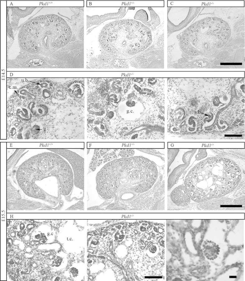
Hematoxylin and Eosin analysis of kidneys. (A–C) Hematoxylin-Eosin staining of Pkd1+/+, Pkd1+/− and Pkd1−/− kidneys at day E14.5 shows no major developmental defects of Pkd1−/− kidneys. Bar = 500 μm. (D) Condensed mesenchyme (cm) surrounding the ureteric bud (ub) as well as more advanced structures such as coma-shaped and s-shaped bodies (ssb) can be identified in Pkd1−/− kidneys. Some rare glomerular cysts are already present (gc). Bar = 500 μm. (E–G) Hematoxylin-Eosin staining of kidneys Pkd1+/+, Pkd1+/− and Pkd1−/− at day E15.5 shows formation of both tubular and glomerular cysts in Pkd1−/− kidneys. Bar = 500 μm. (H) Epithelializing structures and both glomerular (gc) and tubular cysts (tc) are visible in Pkd1−/− kidneys. Bar = 100 μm on the left, 20 μm on the right.