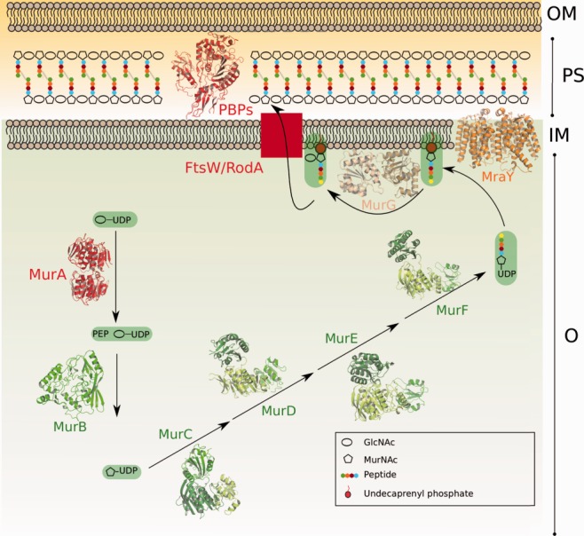
Figure 1.

Schematic diagram of the cytoplasmic and membrane steps of the peptidoglycan biosynthetic pathway. The different domains of Mur enzymes are shown in shades of green. MurA and PBPs, which are the targets of antibiotics currently employed in hospital settings, are highlighted in red. OM, outer membrane; PS, periplasm; IM, inner membrane; C, cytoplasm.
