
Figure 4.
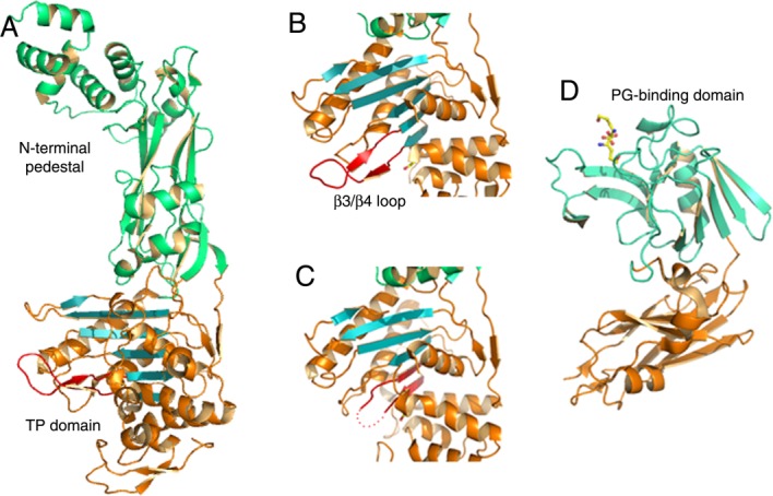
PBPs and l,d-transpeptidases recognize peptidoglycan and β-lactams through α/β folds. (A) PBP2b from S. pneumoniae folds into distinct domains, where the C-terminal, transpeptidase domain harbors the active site within an α/β fold, (B) Zoom of the β3/β4 region of PBP2b (indicated in red), which shows flexibility in a number of PBPs and peptidoglycan-recognizing enzymes. (C) Same region as in (B), but from PBP2b from a drug-resistant S. pneumoniae strain, indicating that the loop between β3/β4 could not be traced in the electron density map and is thus indicated with dots. (D) Structure of l,d transpeptidase from M. tuberculosis bound to a short region of a peptidoglycan substrate.