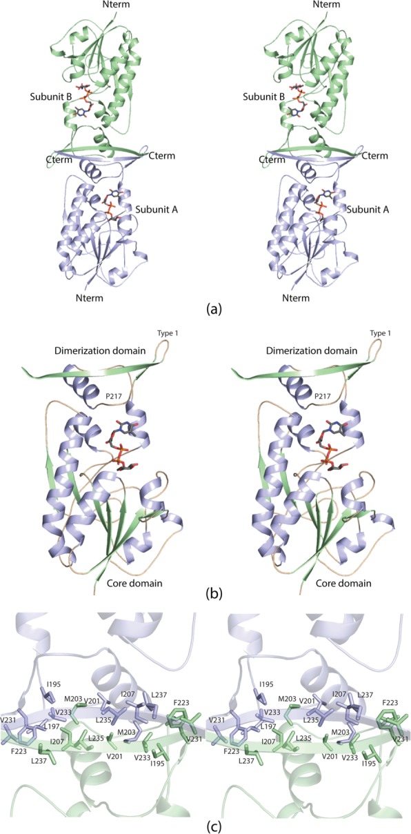
Figure 3.

The structure of WbtJ. A ribbon representation of the WbtJ dimer is displayed in (a). The subunit:subunit interface is formed by a four stranded β-sheet. The dTDP-sugar ligands are displayed as sticks. Shown in (b) is a ribbon representation of one subunit. The overall architecture of the subunit can be envisioned as a globular “core” domain that harbors the active site region and a dimerization domain that extends away from the core motif. A close-up view of the subunit:subunit interface is presented in (c). Subunits A and B are colored in light blue and green, respectively.
