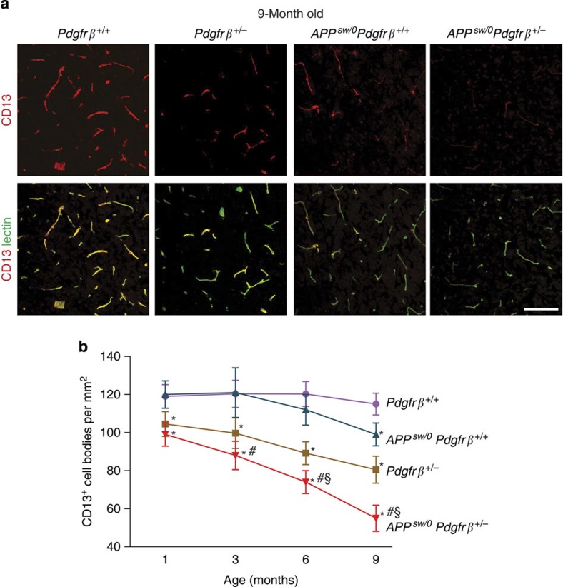
Figure 1. Progressive degeneration of pericytes in APPsw/0Pdgfrβ+/− mice.
(a) Confocal microscopy analysis of CD13-positive pericytes and lectin-positive capillary endothelium in the cortex of 9-month-old Pdgfrβ+/+, Pdgfrβ+/−, APPsw/0; Pdgfrβ+/+ and APPsw/0; Pdgfrβ+/− mice. Scale bar, 100 μm. (b) Quantification of CD13-positive pericytes in the cortex and hippocampus of 1-, 3-, 6- and 9-month-old Pdgfrβ+/+, Pdgfrβ+/−, APPsw/0; Pdgfrβ+/+ and APPsw/0; Pdgfrβ+/− age-matched littermates. Mean±s.e.m., n=6 mice per group. Data from the cortex and hippocampus were pooled because there were no significant differences between these two regions. *P<0.05, all other groups compared with Pdgfrβ+/+; #P<0.05, APPsw/0; Pdgfrβ+/− compared with APPsw/0; Pdgfrβ+/+; §P<0.05, APPsw/0; Pdgfrβ+/− compared with Pdgfrβ+/−. All comparisons are by analysis of variance (ANOVA) followed by Tukey’s post-hoc tests.

