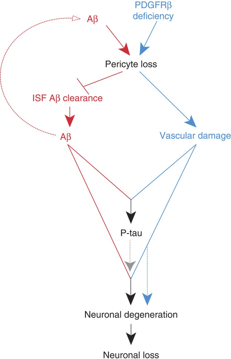
Figure 10. Pericyte loss influences multiple steps of Alzheimer’s-like pathogenic cascade in APPsw/0 mice.
Within the Aβ pathway (red), pericyte loss in APPsw/0 mice because of PDGFRβ deficiency and/or excessive accumulation of Aβ in pericytes diminishes early in the disease process clearance of soluble Aβ from brain ISF causing an early Aβ accumulation and deposition in the brain, which in turn amplifies the loss of pericytes. Within the vasculature, an Aβ-independent pathway driven by aberrant PDGFRβ signalling in pericytes (blue) leads to accelerated pericyte loss that amplifies blood–brain barrier disruption and microvascular reductions in APPsw/0 mice and the degree of vascular damage. Both pathways acting in parallel lead to an early development of a full spectrum of AD-like pathology in mice including significant Aβ pathology, tau pathology, neuronal degeneration and neuronal loss that are not observed either within the Aβ pathway alone or the vascular Aβ-independent pathway alone.

