Figure 5.
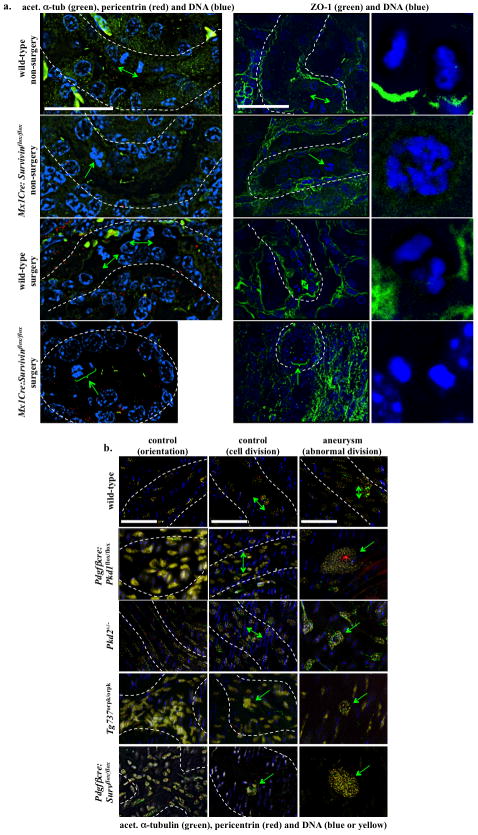
Abnormal cellular division orientation is associated with renal cystic and vascular aneurysm phenotypes. (a) Kidney tubular sections from both Mx1Cre:survivinflox/flox mice, with or without UUO surgery, showed abnormal cell division and division orientation with respect to the axis of the kidney tubule. ZO-1 staining was used to indicate renal tubule orientation, and cell-undergoing division within the region is further enlarged. (b) Longitudinal abdominal aortic sections in non-surgery (control) and aneurysm-induced (surgery) models were studied to analyze endothelial orientation and cell division. Nucleus from smooth muscle cells was shown in blue, and nucleus of a single intimal layer of endothelial tissue was pseudo-colored in yellow. Abnormal randomized cell orientation is clearly visible. In all figures, division orientation relative to tubule/artery axis is shown in green double head arrows, and abnormal cell division is indicated in green arrows. N≥3 for each group and genotype. N≥100 for distribution of spindle orientation angle for each genotype and each treatment. Bar=40 μm.

