Figure 6.
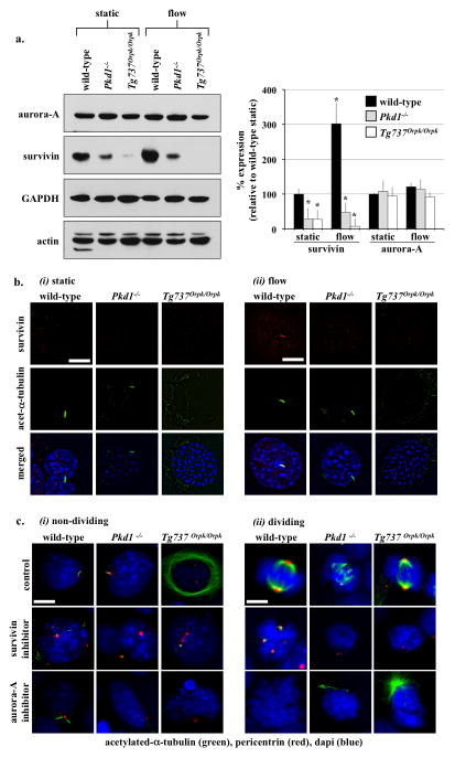
Cilia regulate cell division through survivin expression. (a) Western blot analysis was used to study survivin and aurora-A expressions in wild-type and cilia mutant cells (Pkd1−/− and Tg737Orpk/Orpk) in the presence or absence of fluid-shear stress. GAPDH and actin were used as loading controls. Bar graph represents averaged survivin and aurora-A expressions. (b) Acetylated-α-tubulin was used as a ciliary marker to indicate ciliary expression and localization of survivin in response to fluid-shear in wild-type but not mutant cells. (c) Cells treated with survivin or aurora-A inhibitors are characterized by multiple centrosomes, abnormal mitotic spindle, and mitotic arrest during cell division. Cells were stained with acetylated-α-tubulin (green) and pericentrin (red) and captured at resting (i) and dividing (ii) stages of cell cycle. Bar=10 μm. N=3 for each cell type and treatment. Statistics was performed by comparing individual group to their corresponding wild-type static control groups.

