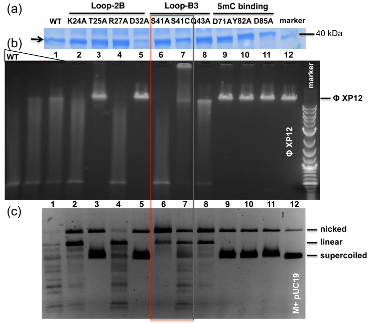
Figure 4. AspBHI variants and activity assays on modified plasmid and phage DNA substrates.
(a) SDS-PAGE analysis of partially purified His-tagged AspBHI WT and its variants after nickel-chelated affinity chromatography. Arrow indicates the AspBHI protein band. (b) Endonuclease activity assay on phage XP12 DNA containing 5mC. Three concentration of WT AspBHI (~0.57 pmoles, with 2-fold serial dilution) were used in the digestion. Mutant enzyme concentrations were estimated at 0.29 to 0.57 pmoles. The smearing may result from partial digestions of the phage DNA. We note that S41C protein tends to precipitate in conditions with <0.2 M NaCl. (c) Endonuclease activity assay on Dcm+ and M.HpaII modified pUC19 DNA.

