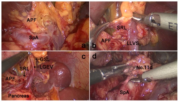
Figure 2. Dissection of the No.

a. The APF was completely lifted cephalad to fully expose the superior border of the pancreas and enter the retropancreatic space (RPS). b.The lower lobar vessels of the spleen (LLVSs) were exposed between the two layers of the SRL by following the RPS c. The left gastroepiploic vessels were exposed by following the fascias. d. Dissection of the No. 11d LN was performed by following the intrafascial space. APF: Anterior pancreatic fascia; SRL, Splenorenal ligament; LLVSs, Lower lobar vessels of spleen; GSL, Gastrosplenicligament; LGEVs, Left gastroepiploic vessels; SPA, Splenic artery.
