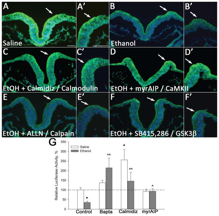
Figure 5. CaMKII inhibition but not other β-Catenin effectors stabilize β-catenin protein.
(A–F) Neural folds of 3-somite embryos were treated with the indicated inhibitor prior to 52mM ethanol exposure; β-catenin was visualized 2hr thereafter in presumptive hindbrain. Arrows indicate the neural crest-enriched dorsal neural fold; scale bar represents 50 μm. Saline control (A) has robust β-catenin distribution within the dorsal neuroepithelium (arrow), and this is substantially reduced by ethanol (B). The calmodulin-selective inhibitor calmidizolium (C) (20 μM) and the CaMKII-selective inhibitor myrAIP (D) (5 μM) both prevent β-catenin loss within the ethanol-treated dorsal neural folds. In contrast, inhibitors directed against calpain (E, ALLN, 5 μM) and GSK-3β (F, SB-415,286, 5 μM) do not prevent ethanol-mediated β-catenin loss. (G) CaMKII and Cai2+ inhibition restore β-catenin transcriptional activity in ethanol-treated hindbrain. Hindbrain expression of TopFlash luciferase reporter was quantified and normalized against Renilla luciferase co-expression in hindbrains pretreated as indicated prior to saline or ethanol challenge. Mean ± SEM of three experiments having 6–8 embryos/treatment, normalized to saline control. * p < 0.001 vs. saline-only; ^ p < 0.05 and ^^ p < 0.001 vs. ethanol-only, analyzed using one-way ANOVA with Holm-Sidak method.

