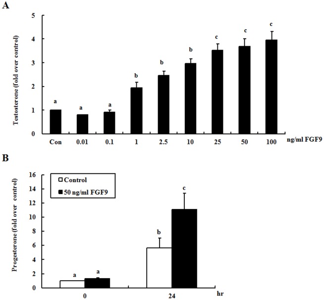
Figure 1. FGF9 increases steroidogenesis in both mouse primary and tumor Leydig cells.
(A) Mouse primary Leydig cells were treated with different concentrations of FGF9 for 24 hr (Con = control). (B) MA-10 mouse Leydig tumor cells were treated without (control) or with 50 ng/ml FGF9 for 0 and 24 hr. At the end of each incubation, culture medium was collected, and testosterone (A) or progesterone (B) production was measured by RIA. Each bar represents the mean ± SEM of the fold difference in testosterone or progesterone production compared with the control group. Different letters above the bars indicate significant differences between treatments (p<0.05).

