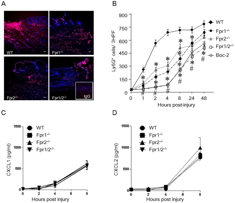
Figure 2. Reduced neutrophil infiltration in the wounds of Fpr-deficient mice and the production of chemokines.
A, representative immunofluorescence of skin wound showing Ly6G+ cells 4 h after injury. Cryosections of wounded skin from WT and Fpr-deficient mice were labeled with Ly6G and DAPI (Red: Ly6G; Blue: DAPI) (n = 5, scale bar: 20 µm). Insert: control IgG staining. B, Kinetics of infiltrating Ly6G+ neutrophils in 3 consecutive high power fields (HPF). *, significantly reduced Ly6G+ cells in the wounds of Fpr-deficient micecompared with WT mice (p< 0.05). #, significantly reduced Ly6G+ cells in the wounds of Boc-2 pretreated WT mice, compared with WT mice without pretreatment (p< 0.05). C&D, chemokinesCXCL1 and CXCL2 in the homogenates of skin wound from WT and Fpr-deficient mice. Skin (1 cm×1 cm) with wounds in the center was excised and homogenized in 5 ml DPBS. The homogenates were centrifuged and the supernatants were collected for measurement of CXCL1 and CXCL2 by ELISA (n = 15).

